
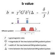
B γ² G² δ² Δδ3 Therefore a larger b value is achieved by increasing the gradient amplitude and duration and by widening the interval between paired. With enhanced gradients the whole b ra in can b e scanned with in seconds.
The Basics Of Mri Interpretation Radiology Geeky Medics
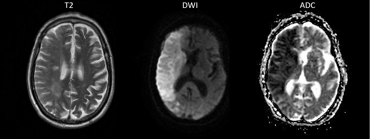
DWI is done to determine the rate of molecular diffusion in different areas of the body.
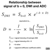
. B value measures the degree of diffusion weighting applied thereby indicating the amplitude G time of applied gradients δ and duration between the paired gradients Δ and is calculated as. Our purpose was to evaluate the appearance of the normal brain on DW MR images as the diffusion gradient strength b value is increased from 1000 to 3000 smm2. The best b-value combination was 0 and 600 smm2 and multiple b2.
Multiple b values of 600 smm2 and higher are recommended to differentiate between benign and malignant. Using a Gaussian model with b-values up to 4000 smm 2 Mardor et al. The b value is a parameter that is used in diffusion weighted imaging.
The strong correlation implies that tumors with low. The aim of this work is to determine the role of diffusion-weighted magnetic resonance imaging DW-MRI and the apparent diffusion coefficient ADC in the differentiation between benign and malignant solid head and. A baseline b-value of 50 smm² is often used in liver diffusion-weighted imaging instead of b 0.
Differentiation between malignant and benign masses is essential for treatment planning and helps in improving the prognosis of malignant tumors. In general in healthy tissue molecules of water and other chemicals are not stationary but moving about. B γ² G² δ² Δδ3 Therefore a larger b value is achieved by increasing the gradient amplitude and duration and by widening the interval.
In general approximately 1000 smm 2 is the maximal b value for DWI 5 11. The reason is readily apparent from the images below. National Center for Biotechnology Information.
MRI protocol included 3DT2w images high resolution Fov Optimized and Constrained Undistorted Single-Shot FOCUS DWI images with b-values of 100 400 800 and 2000 smm 2 and dynamic contrast enhanced images. B value measures the degree of diffusion weighting applied thereby indicating the amplitude G time of applied gradients δ and duration between the paired gradients Δ and is calculated as. Typical b-values available on modern MRI scanners range from 0 to about 4000 smm².
The lesionnormal parenchymal ADC ratio for b600 b1000 and multiple b2 better distinguished between benign and malignant lesions. The purpose of the bipolar gradient is to force a phase shift in our tissues. The higher the b values the better the sensitivity of diffusion weighted imaging usually three to four b values are used in diffussion-weighted sequences b50 b500 b1000 and b1400.
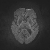
In DWI the optimal b value is 600 smm2. Brain DW images obtained at b 3000 appear significantly different from those obtained at b 1000 reflecting expected loss of signal from all areas of brain in proportion to their ADC values. Tissues that are free moving and unrestricted isotropic diffusion will produce a.
High b-values with or without non-Gaussian models have been used for early evaluation of cancer treatment. C-DWI images 2000 and 2500 smm 2 were derived from the three lower acquired b-value DWI images using a mono-exponential diffusion decay. The term b -value derives from the landmark 1965 paper by Stejskal and Tanner in which they described their pulsed gradient diffusion method.
The b value is used in MRI in the context of Diffusion Weighted Imaging DWI. Reported that pre-treatment ADC and a diffusion index RD can both correlation well r 076077 with brain tumor response to radiation therapy 87. In DWI we recommend the use of b-values of 0 and 800 smm2 as two b-values or b0 50 600 800 and 1000 smm2 as multiple b-values for distinguishing between benign and malignant liver lesions.
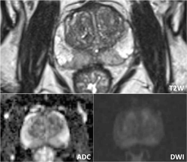
Diffusion-weighted MRI and optimal b-value for characterization of liver lesions. Strength duration and the period b etween diffusion gradients. Beside the diffusion-weighted row data images there is one more way to present diffusion data which is called the apparent diffusion coefficient ADC map of water.
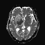
The degree of diffusion weight in g correlates with the strength of the diffusion gradients characterized b y the b - value which is a function of the gradient related parameters. It controls the amplitudestrength of the bipolar gradient coils activated to detect thermal motion of water in and around cells Brownian motion. With b 0 bright signals are noted in multiple veins due to the high T2 of blood coupled with sluggish flow.
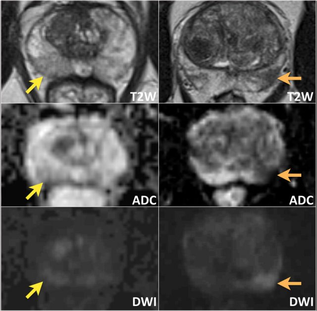
Studies have reported that the use of b values higher than 1000 smm 2 and 2000 smm 2 improves tumor localization and the contrast between benign and malignant lesions in the prostate and the breasts 12 14.

Principles Of Diffusion Tensor Imaging And Its Applications To Basic Neuroscience Research Neuron
Diffusion Weighted Imaging Radiology Reference Article Radiopaedia Org
Apparent Diffusion Coefficient Radiology Reference Article Radiopaedia Org
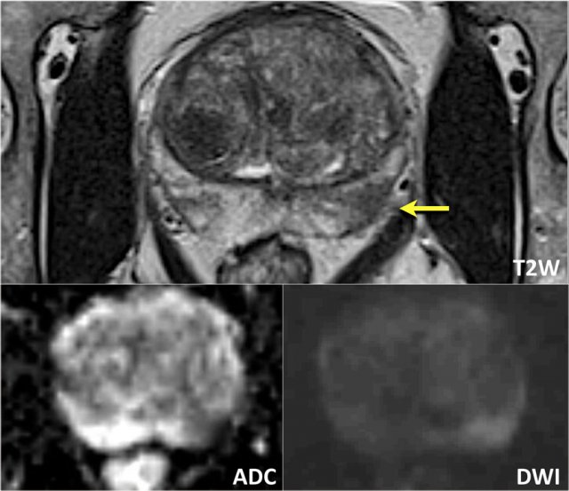
The Radiology Assistant Prostate Cancer Pi Rads V2
Diffusion Weighted Imaging Radiology Reference Article Radiopaedia Org
Diffusion Weighted Imaging Radiology Reference Article Radiopaedia Org
The Radiology Assistant Prostate Cancer Pi Rads V2

Diffusion Tensor Imaging Dti Fiber Tracking Imagilys
The Radiology Assistant Prostate Cancer Pi Rads V2
Tensor Valued Diffusion Encoding For Diffusional Variance Decomposition Divide Technical Feasibility In Clinical Mri Systems Plos One
Brain Segmentation Software Quantib Neurodegenerative Quantib Nd

Apparent Diffusion Coefficient Radiology Reference Article Radiopaedia Org

Apparent Diffusion Coefficient Radiology Reference Article Radiopaedia Org

Hyperpolarised 13c Mri Identifies The Emergence Of A Glycolytic Cell Population Within Intermediate Risk Human Prostate Cancer Nature Communications

Anwar Padhani Profpadhani Twitter
Tensor Valued Diffusion Encoding For Diffusional Variance Decomposition Divide Technical Feasibility In Clinical Mri Systems Plos One
2

Diffusion Weighted Imaging Radiology Reference Article Radiopaedia Org

Diffusion Tensor Imaging Dti Fiber Tracking Imagilys